
Member-only story
Endoscopic Anatomy of the Paranasal Sinuses: A Comprehensive Guide

Mike Hayes
·12.5k Followers· Follow Published in Endoscopic Anatomy Of The Paranasal Sinuses
5 min read 969 View Claps
58 Respond
The paranasal sinuses are a group of air-filled cavities located within the facial bones. They are lined with mucous membranes and help to warm, moisten, and filter the air we breathe. The sinuses also help to produce mucus, which helps to protect the nasal passages from infection. The paranasal sinuses are divided into four pairs: * **Frontal sinuses** are located in the forehead. * **Maxillary sinuses** are located in the cheekbones. * **Ethmoid sinuses** are located between the eyes. * **Sphenoid sinuses** are located behind the nose. The paranasal sinuses are connected to the nasal cavity by small openings called ostia. These ostia allow air and mucus to flow in and out of the sinuses. <h2>Endoscopic Anatomy</h2> Endoscopy is a medical procedure that uses a thin, flexible tube with a camera on the end to visualize the inside of the body. Endoscopy can be used to examine the paranasal sinuses and to diagnose and treat sinus infections and other conditions. During an endoscopic examination of the paranasal sinuses, the endoscope is inserted into the nasal cavity and advanced into the sinuses. The camera on the end of the endoscope allows the doctor to see the inside of the sinuses and to identify any abnormalities. The endoscopic anatomy of the paranasal sinuses is complex and varies from person to person. However, there are some general features that are common to all sinuses. The **frontal sinuses** are located in the forehead and are the largest of the paranasal sinuses. They are divided into two parts by a thin bony septum. The **maxillary sinuses** are located in the cheekbones and are the second largest of the paranasal sinuses. They are also divided into two parts by a thin bony septum. The **ethmoid sinuses** are located between the eyes and are the smallest of the paranasal sinuses. They are made up of a complex network of thin bony plates. The **sphenoid sinuses** are located behind the nose and are the deepest of the paranasal sinuses. They are also made up of a complex network of thin bony plates. The **ostia** of the paranasal sinuses are located in the nasal cavity. The **ostium** of the frontal sinus is located in the middle meatus of the nose. The **ostium** of the maxillary sinus is located in the inferior meatus of the nose. The **ostia** of the ethmoid sinuses are located in the superior meatus of the nose. The **ostium** of the sphenoid sinus is located in the sphenoethmoidal recess of the nose. <h2>Clinical Significance</h2> The endoscopic anatomy of the paranasal sinuses is important for a number of reasons. First, it allows doctors to visualize the inside of the sinuses and to identify any abnormalities. This information can be used to diagnose and treat sinus infections and other conditions. Second, the endoscopic anatomy of the paranasal sinuses can be used to guide surgical procedures. For example, endoscopic surgery can be used to open blocked ostia, to remove polyps, and to treat other sinus conditions. The endoscopic anatomy of the paranasal sinuses is a complex and important topic. A thorough understanding of the endoscopic anatomy of the paranasal sinuses is essential for the diagnosis and treatment of sinus infections and other conditions. <h2>References</h2> * [1] Stammberger, H., & Wolf, G. (2000). Endoscopic anatomy of the paranasal sinuses. GMS current topics in otolaryngology-head and neck surgery, 4(2). * [2] Bolger, W. E., Zinreich, S. J., & Hawke, M. (2003). Endoscopic sinus surgery: An anatomical and surgical approach. Plural Publishing. * [3] Kennedy, D. W., & Zinreich, S. J. (2010). Functional endoscopic sinus surgery: The endoscopic approach to the paranasal sinuses and skull base. Thieme.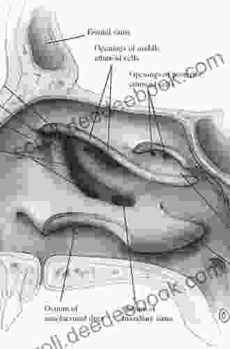
Endoscopic Anatomy of the Paranasal Sinuses
by Peter S. Hechl
4.6 out of 5
| Language | : | English |
| File size | : | 45278 KB |
| Text-to-Speech | : | Enabled |
| Enhanced typesetting | : | Enabled |
| Print length | : | 150 pages |
| Screen Reader | : | Supported |
Create an account to read the full story.
The author made this story available to Deedee Book members only.
If you’re new to Deedee Book, create a new account to read this story on us.
Already have an account? Sign in
969 View Claps
58 Respond
Join to Community
Do you want to contribute by writing guest posts on this blog?
Please contact us and send us a resume of previous articles that you have written.
Resources
 Book
Book Page
Page Chapter
 Text
Text Reader
 Library
 Paperback
 E-book
 Magazine
 Paragraph
 Bookmark
 Shelf
Shelf Glossary
 Bibliography
 Preface
 Synopsis
 Codex
Codex Tome
Tome Classics
 Narrative
 Autobiography
 Memoir
 Reference
 Thesaurus
 Narrator
 Character
 Resolution
 Catalog
 Study
Study Lending
 Academic
 Reading Room
 Rare Books
 Special Collections
 Literacy
 Thesis
 Awards
 Reading List
 Book Club
 Theory
 Amanda Wills
 Michael Rectenwald
 Donatella Mutolo
 Katie Hornor
 Candace Cotton
 William Cowper
 Melvin M Belli
 Amanda Lovelace
 Simon Hepworth
 Kelly Grant
 Antoinette Portis
 John Lars Shoberg
 R Bick Lesser
 Stephen Michael Shearer
 Karyne E Messina
 Leckie
 James Montague
 Frank Smyth
 Dan Grec
 Nikki Landis
Light bulbAdvertise smarter! Our strategic ad space ensures maximum exposure. Reserve your spot today!


Good Author
 Melvin BlairFollow ·6.3k
 Colton CarterFollow ·15.1k
 Jacob HayesFollow ·16.7k
 Preston SimmonsFollow ·11.1k
 Grant HayesFollow ·2.5k
 Charles BukowskiFollow ·19.9k
 Terry BellFollow ·10.9k
 Brandon CoxFollow ·11.7k
Recommended from Deedee Book

 Howard Powell
Dk Workbooks Science Third Grade: An In-Depth Exploration...
Science education plays a...
·4 min read
1.5k View Claps
89 Respond

 José Saramago
·6 min read
1.3k View Claps
74 Respond

 Everett Bell
Learn to Play Bluegrass Dobro Guitar: A Comprehensive...
: Bluegrass Dobro, A Story of...
·5 min read
872 View Claps
60 Respond

 Jeffrey Cox
How the Raccoon Got His Mask
The raccoon, with its...
·3 min read
144 View Claps
18 Respond

 George Bell
Hannah Meets Ruby Hannah Out West: An Adventure-Filled...
Hannah Meets...
·4 min read
66 View Claps
7 Respond

 Wade Cox
·6 min read
686 View Claps
41 Respond
The book was found!
Endoscopic Anatomy of the Paranasal Sinuses
by Peter S. Hechl
4.6 out of 5
| Language | : | English |
| File size | : | 45278 KB |
| Text-to-Speech | : | Enabled |
| Enhanced typesetting | : | Enabled |
| Print length | : | 150 pages |
| Screen Reader | : | Supported |
